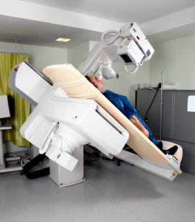
 Fluoroscopy is the term used for continuous, or moving, x-ray imaging. If you think of an x-ray as a digital photo, fluoroscopy is like a digital movie. The technique of fluoroscopy is used for a series of exams, mostly used to evaluate the GI system (esophagus, stomach, bowel, and colon) or the genitourinary system (kidneys, ureters, and bladder). For GI tests, fluoroscopy uses barium as a contrast agent, either after drinking it or after administering through a tube in the rectum. For fluoroscopic exams of the bladder, contrast is gently used through a Foley catheter. Types of studies that use fluoroscopy include esophogram, upper GI series, small bowel series, barium enema, VCUG, and cystography.
Excessive exposure to radiation can be harmful, but the exposure during a fluoroscopy exam is generally minimal and the benefits of an accurate diagnosis far outweigh the risk. Moreover, we use every safety precaution to use the least possible amount of radiation, especially for children. It is important to inform your doctor of any recent illnesses or medical conditions as some conditions may increase the risk of an adverse effect.
Women should always inform their physician and the technologist if they are pregnant or may be pregnant. Nursing mothers should wait 24 hours after receiving IV contrast before resuming breast-feeding. Allergic reaction to contrast (mild or severe) is also a rare but possible risk. Our team is well-prepared to deal with such reactions should the need arise.
Fluoroscopic imaging procedures performed by our experienced staff: